New CT Scanner Opens New Views into Fish Evolution at Tulane


Rows of preserved fishes line the shelves inside Tulane University's Biodiversity Research Institute. Some differ in obvious ways. Others appear almost indistinguishable, even to trained eyes. For PhD candidate Godservice Eziefule, who goes by GS, those subtle differences are exactly what make them fascinating. Hidden inside each specimen are clues to millions of years of evolution waiting to be uncovered.
Until recently, getting a detailed look at those clues could require traveling hundreds or even thousands of miles to reach specialized imaging equipment. GS has traveled twice to Houston, Texas, and once to Friday Harbor, Washington, to scan specimens for his research.
Now, much of that work can happen at Tulane.
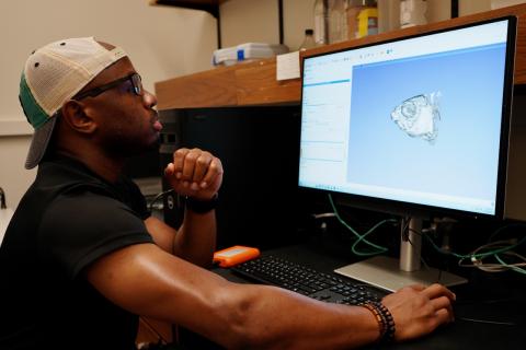
A new Bruker SKYSCAN 1273 micro-CT scanner in Tulane's Department of Ecology and Evolutionary Biology allows researchers to create detailed three-dimensional images of the internal structures of preserved specimens without cutting them apart. For researchers studying how fishes differ, how they evolved, and even whether similar-looking specimens represent different species, those images can reveal an entirely new layer of information.
For GS, who came to Tulane after training in fisheries with a focus on conservation biology and ecology in Nigeria, that means looking beneath the exterior of West African fishes and closely examining their skeletons. His research uses differences in skull anatomy to investigate evolutionary relationships among fishes that can be difficult to distinguish through external features alone.
One structure of particular interest is the kinethmoid, a small bone involved in the feeding mechanics of minnows, carps and their close relatives. By comparing its shape and other skeletal characteristics across specimens, GS can investigate how different forms may relate to evolutionary history.
The work connects back to the questions that first drew him to fish research: understanding biodiversity well enough to help conserve it. Knowing what species exist, how they differ, and how they are related provides information scientists can use to better understand the ecosystems in which those species live.
The scanner dramatically changes how researchers can pursue those questions.
Brian Sidlauskas, director and curator of the Tulane University Biodiversity Research Institute and Royal D. Suttkus Fish Collection and professor of ecology and evolutionary biology, said access to micro-CT technology was one of the factors that convinced him to leave Oregon State University and come to Tulane.
At his previous institution, Sidlauskas did not have ready access to a scanner for his lab. When he needed one, he sometimes traveled hours to another facility and worked around whatever scanner time was available. At Tulane, the technology is part of the research environment he and his students use every day.
For Sidlauskas, that access is increasingly essential to modern evolutionary biology. Genetic information can help scientists understand how organisms are related, but genetics alone does not explain how those organisms differ physically, how they interact with their environments or how their forms have changed through evolutionary time.
“The phenotype adds all of the context to the genetics,” Sidlauskas said. “With just the genotype you can generate an evolutionary tree and say these are the branches. But to add all of the color and to add all of the understanding of what those relationships imply, you need the phenotype as well.”
Micro-CT imaging allows researchers to take that investigation inside the animal, examining structures that would otherwise require extensive preparation or dissection. Sidlauskas said the technology has transformed his field over the past two decades, giving researchers ways to study preserved specimens that were once extraordinarily difficult.
GS's research provides a particularly striking example.
Sidlauskas said the large size of the fishes GS studies would make some traditional methods of skeletal preparation impractical. Producing the skulls needed for his research could require extensive dissection and preparation of fresh specimens. Instead, a preserved fish can be scanned and transformed into a digital three-dimensional model of its skeleton.
“I don't think that GS's project actually would be possible without it,” Sidlauskas said. “Here, he just sticks the fish in the scanner, waits a few hours, and he has a model.”
Sidlauskas knows the difference firsthand. During his own doctoral research roughly 25 years ago, Sidlauskas studied much smaller fish, like those Basil Williams, one of his current PhD students, is studying today. He spent months preparing tiny skeletons, dissecting them into individual components and moving specimens one by one beneath a microscope to compare bones and record differences.
“I would have finished my PhD a whole year earlier if I had access to this technology, possibly more than that,” Sidlauskas said.
Williams is now using that technological advantage to investigate a different evolutionary puzzle.
A second-year PhD student who previously worked as an assistant collection manager at the Florida Museum of Natural History, Williams studies Enteromius, a widespread group of small African barbs. Her research combines taxonomy with ecomorphology, examining how factors such as diet and habitat relate to skeletal structure and body shape.
The fish present a fundamental problem for a taxonomist: many of them look remarkably similar.
“My fish are very hard to tell apart,” Williams said. “They're just minnows. They just look like silver fish.”
Genetic and biogeographic evidence suggests there may be undescribed species among the populations she studies. Williams is looking inside the fish for anatomical differences that could help distinguish them, and the scans are already revealing promising clues.
After scanning dozens of specimens, Williams began seeing differences in their skulls. In two populations, structures around the eye sockets appeared distinctly different.
“I was like, ‘Yes, thank you,’” Williams said.
Seeing a difference, however, is only the beginning. Williams uses a process called landmarking to place consistent reference points across the three-dimensional skull models. She can then mathematically compare the positions of those points, turning observations such as one skull appearing longer and narrower than another into quantitative measurements that can be statistically analyzed.
That scale of analysis is possible in part because the scanner is not located at a distant shared facility.
At some institutions, researchers may wait weeks or months for limited blocks of scanner time. At Tulane, students in Sidlauskas' lab can use the equipment regularly, allowing them to scan more specimens and pursue larger data sets. Williams estimates that her dissertation research could eventually involve hundreds of scans.
The experience is also becoming part of students' scientific training. Williams, who learned CT scanning before arriving at Tulane, is now helping train others. During her interview for this story, she was preparing to train a first-year undergraduate researcher who was only weeks into her Tulane education.
That accessibility is already prompting researchers to imagine uses beyond the projects for which the scanner was originally acquired. Students have approached the lab with ideas ranging from examining plant root structures to studying brains and braincases. Sidlauskas said the technology could also support research involving biological materials, archaeological objects, engineered materials, and any other specimens that can fit inside the instrument.
But perhaps its greatest potential lies in what is already in Tulane's care.
The Royal D. Suttkus Fish Collection contains more than 7 million preserved fish specimens representing approximately 3,800 species. Built originally through more than five decades of fieldwork led by renowned Tulane ichthyologist Royal D. Suttkus and augmented by thirty-five years of work by Hank Bart (now director emeritus), the collection is particularly rich in fishes from the Gulf South and includes extensive sampling of regional waterways over time. Those specimens form a biological archive that researchers can now revisit using technologies that did not exist when many of them were collected.
“The collection is an enormous resource, and this unlocks things from the collection that we couldn't do with it ahead of time,” Sidlauskas said.
Over time, he hopes the lab can create a growing virtual library by scanning representative specimens from the collection, with an early emphasis on fishes for which Tulane has particularly valuable examples that may not be well represented elsewhere. Those digital specimens can also be shared with researchers at other institutions and contribute to broader repositories of biological imaging data.
For Williams, whose career interests remain firmly rooted in museum collections, that possibility gets at one of the most exciting things about the technology. Natural history collections may preserve specimens for generations, but new technologies continually create new ways to ask questions of them.
Her description is considerably simpler: scientists keep finding “new things to do with old fish.”
“CT scanning is a very big part of that future for museum collections,” Williams said. “Because we have absurdly easy access now to a really good machine, we can be creative with it.”
That creativity may ultimately be as important as the machine itself.
Sidlauskas does not envision a laboratory in which every student spends years attacking a different piece of a single research question. Instead, he wants students to have the technology, specimens, and scientific support necessary to develop questions of their own. Already, members of the lab are pursuing taxonomy, skeletal anatomy, evolutionary change, hearing and sound production, brain structure, and other areas using many of the same tools.
“It allows my students to be more ambitious,” Sidlauskas said. “It allows us to be more ambitious.”
That philosophy also shapes how he thinks about the future of TUBRI. Sidlauskas has researched questions of his own, including some he hopes to revisit with modern imaging after first encountering them decades ago. But his larger goal is not to build a laboratory around one discovery, and not all of his students study the fishes that he studies.
“I want to build a space in which it's possible for a team of amazing people to answer their own questions rather than simply looking at me to direct everyone,” Sidlauskas said.
The SKYSCAN 1273 adds another powerful tool to that space. Paired with millions of preserved specimens and students eager to ask what else might be hidden inside them, it offers something more valuable than a faster way to answer existing questions.
It creates the opportunity to ask new ones.
For Sidlauskas, that is the legacy he hopes to build at Tulane: not one solved scientific problem, but a collection preserved for future researchers, generations of scientists trained to explore it, and a research environment where discoveries can begin with questions no one has thought to ask yet.